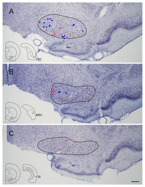
Figure 7.
Distribution of retrogradely labeled cells in HDB following FG injections in LEC (A, case R-27 FG), MEC (B, case R-14 FG), and perirhinal cortex (C, case R-21 FG). Labeled cells are superimposed on the Nissl-stained images of the mapped sections. Blue circles and red rectangles indicate non-cholinergic and cholinergic retrogradely labeled cells, respectively. The injection site and the actual mapped coronal section are shown in the lower left inset. The boxed area on the coronal map shows the position of the picture, and the outline on the Nissl section shows the border of HDB. Notice that the labeled cells are located medially in HDB in the case of MEC injection (B) whereas labeling is present in both medial and ventral portions of HDB in the case of LEC injection (A). In the perirhinal case, labeling is shifted to the mid portion of HDB (C). Scale bar = 0.25 mm.

