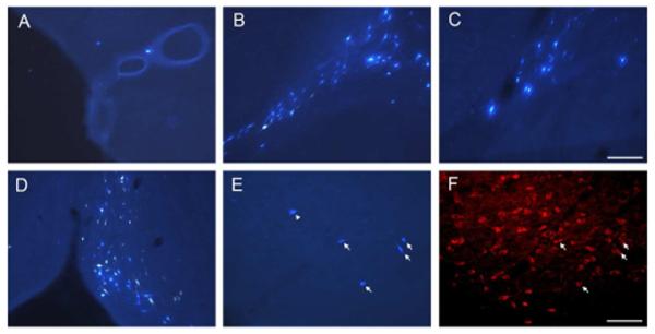
Figure 9.
Photomicrographs of retrogradely labeled cells in the HDB (A) and caudal GP (B) following FB injection in the caudal perirhinal cortex (case R-51 FB), in the caudal GP (C) following FB injection in the postrhinal cortex (case R-29 FB), in the MS/VDB (D) following FB injection in the entorhinal cortex (case R-28 FB). Photomicrographs in E and F show the retrogradely labeled cells in the HDB (E) following FB injection in the caudal perirhinal cortex (case R-50 FB) and immunoreactive cells for ChAT (F) for the same location as in E. Arrows indicate double labeled cells for FB and ChAT (E, F) and an arrowhead indicates a FB labeled cell that is ChAT negative (E). Scale bar = 0.1 mm (A-D) and 0.2 mm (E, F).

