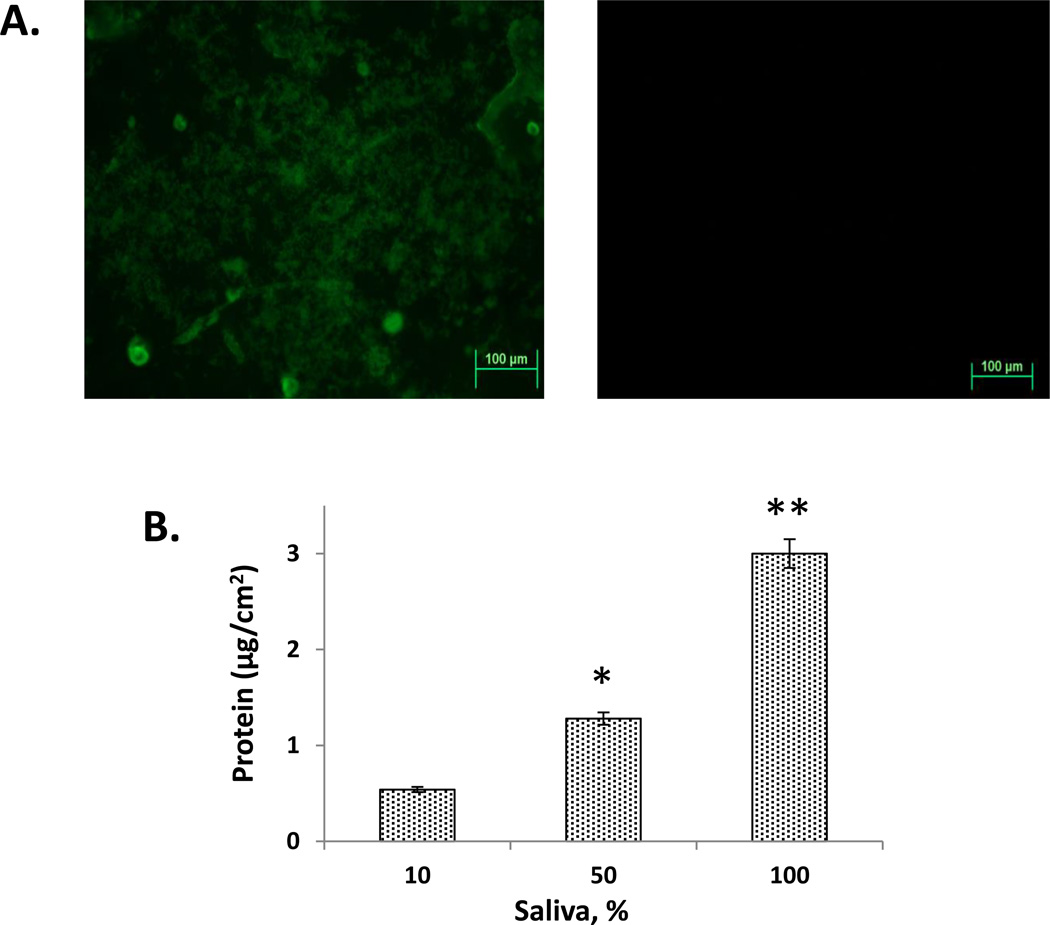
Figure 4. Adsorption of salivary proteins to PMMA-g-PNVP disks.
Panel A: PMMA-g-PNVP disks immersed in 10% human saliva for 20 days were stained with FITC and viewed in a confocal microscope. Positive staining was observed on disks incubated in saliva (left image) but not PBS (right image). Panel B: Quantitation of protein adsorption using the microBCA assay. Disks were incubated for 20 days in 10%, 50%, and 100% saliva, the adsorbed proteins released with SDS, and then assayed. Protein adsorption significantly increased with increasing saliva concentration. *P ≤ 0.015 versus 10%, ** P ≤ 0.001 versus 10%.

