
Figure 2.
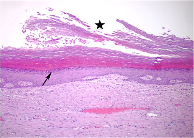
61-year-old male with large left gluteal epidermal inclusion cyst. Representative hematoxalin- and eosin-stained histopathology slide showing the cyst wall lined by stratified squamous epithelium with a granular cell layer (arrow). The central portion of the cyst is filled with keratinaceous debris (star).