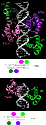
Fig. 8.
3D structure of the POU-specific domain of human OCT1 bound to DNA in two different configurations (Reményi et al. 2001). The HD is in green, and the POU-specific domain is in magenta. Underneath each panel are the respective binding sites used in the X-ray studies as well as schematic views of OCT1 DNA-binding domains. a OCT1 dimer binding to the PORE DNA sequence. The two PAX6 monomers are distinguished by different color intensity (PDB ID: 1HF0). b OCT1 bound to the MORE DNA sequence (PDB ID: 1E30). Note that only half of the dimer is shown, due to the complete symmetry in conjunction with the palindromic binding site.

