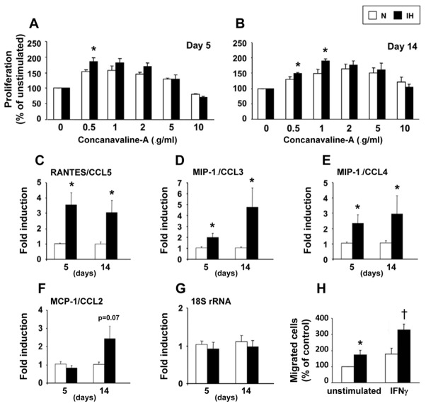
Figure 1. Intermittent hypoxia induces splenocyte activation.
Splenocyte proliferation in response to concanavalin-A after 5 (A) and 14 (B) days of intermittent hypoxia (IH) or air (N); *p<0.05 vs N (n=6–12 per group). Splenocyte mRNA expression of RANTES/CCL5 (C), MIP-1α/CCL3 (D), MIP-1β/CCL4 (E) and MCP-1/CCL2 (F). Measurements were normalized to the eukaryotic 18S ribosomal-RNA (G) and expressed as fold induction of their baseline values; *p<0.05 vs N (n=6 per group). Splenocyte migration toward RANTES/CCL5 after 14 days of IH or air (H). Splenocytes were tested without stimulation or after 50 ng.ml−1 interferon-gamma (IFN-γ) stimulation; p<0.05 vs unstimulated* or stimulated† splenocytes from N-mice (n=4).

