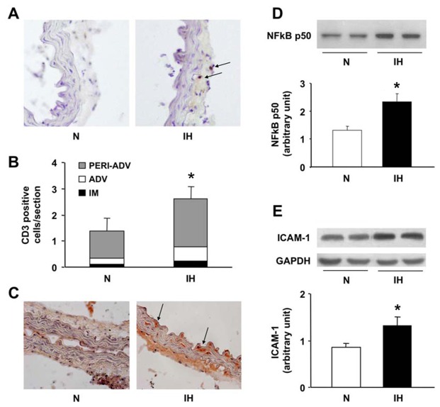
Figure 4. Intermittent hypoxia induces aorta inflammation.
Inflammation was assessed in mice exposed to intermittent hypoxia (IH) or air (N) for 14 days. (A) CD3 immunostaining with arrows showing T-cells (10x20 magnification). (B) Quantitative analysis of CD3-positive cell infiltration according to the various tunica of the aortic wall. Note that T-cells predominated in the adventitia-periadventitia tunica (n=9–10 per group). (C) Representative RANTES/CCL5 immunostaining (10x20 magnification). Immunoblottings and quantifications of nuclear NFkB-p50 (D) and cytosolic ICAM-1 (E) (n=4 each).

