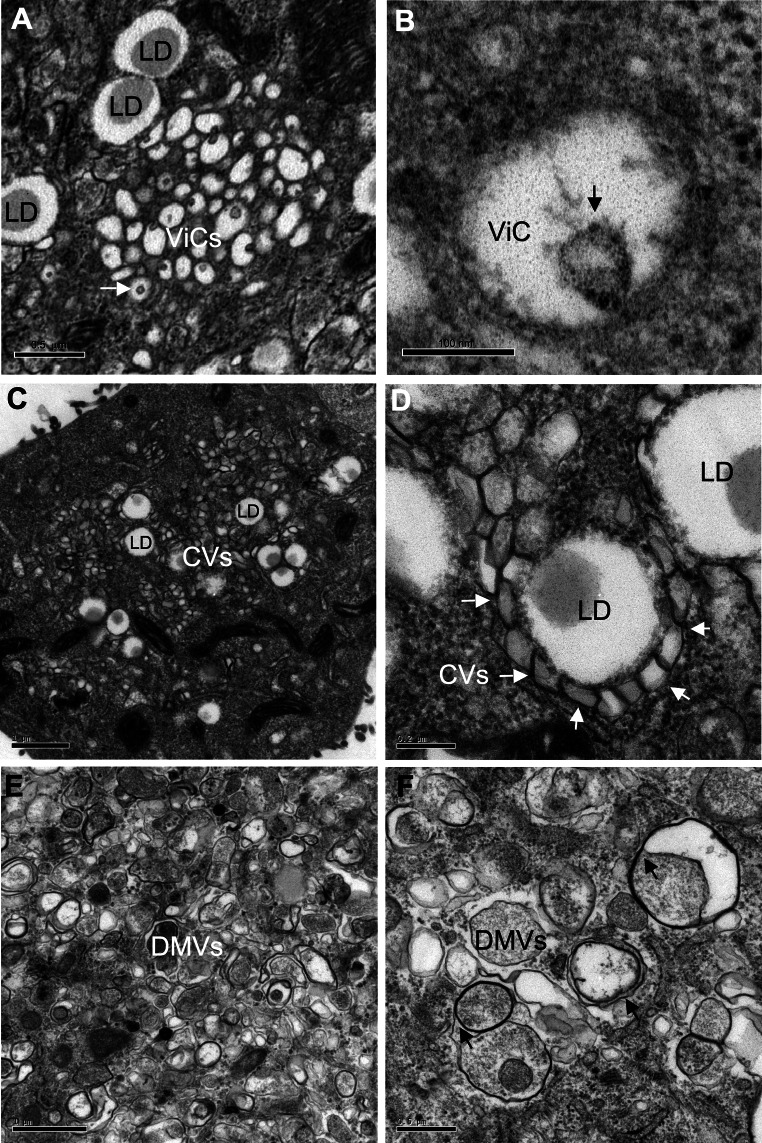
Fig. 1.
Main ultrastructural changes encountered in Huh7.5 cells during the course of HCVcc infection (adapted JFH-1 strain). Cells displayed three types of membrane alterations: a, b vesicles in cluster (ViC), corresponding to a small number (10–40) of single-membrane vesicles of variable size (100–200 nm) grouped together in a well-delimited area; most, if not all, of these vesicles had an internal invagination (white arrow in the low magnification image in a; black arrow in an individual vesicle shown at high magnification in b). c, d Contiguous vesicles (CV), corresponding to single-membrane vesicles of a more homogeneous size (around 100 nm) present in large numbers; these vesicles clustered together and frequently formed a collar around lipid droplets (LD, see the white arrows on the high magnification image in d). e, f Double-membrane vesicles (DMV) of extremely variable size (150–1,000 nm); these vesicles were characterized by a thick, electron-dense membrane consisting of two closely apposed membranes (black arrows on the high magnification image in f). All these structures were highly specific to HCV infection and were not observed in uninfected Huh7.5 cells (not shown)

