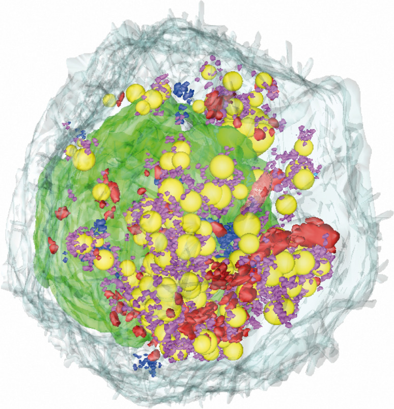
Fig. 4.
Three-dimensional reconstruction of a whole Huh7.5 cell 2 days after infection with HCVcc. The standard EM block was resized for the cutting of a ribbon of 140 serial ultrathin sections (70 nm thick) to reconstruct this particular cell. Contours were drawn with IMOD software through the same specific cellular structures on different serial sections, including the plasma membrane (light gray), the nucleus (green), the lipid droplets (yellow), and the three types of membrane alterations specifically induced by HCV: ViCs (blue), CVs (purple) and DMVs (red). A QuickTime movie of the 3D reconstruction of this cell is also provided as supplemental material, to improve visualization of the spatial distribution of these structures within the cell

