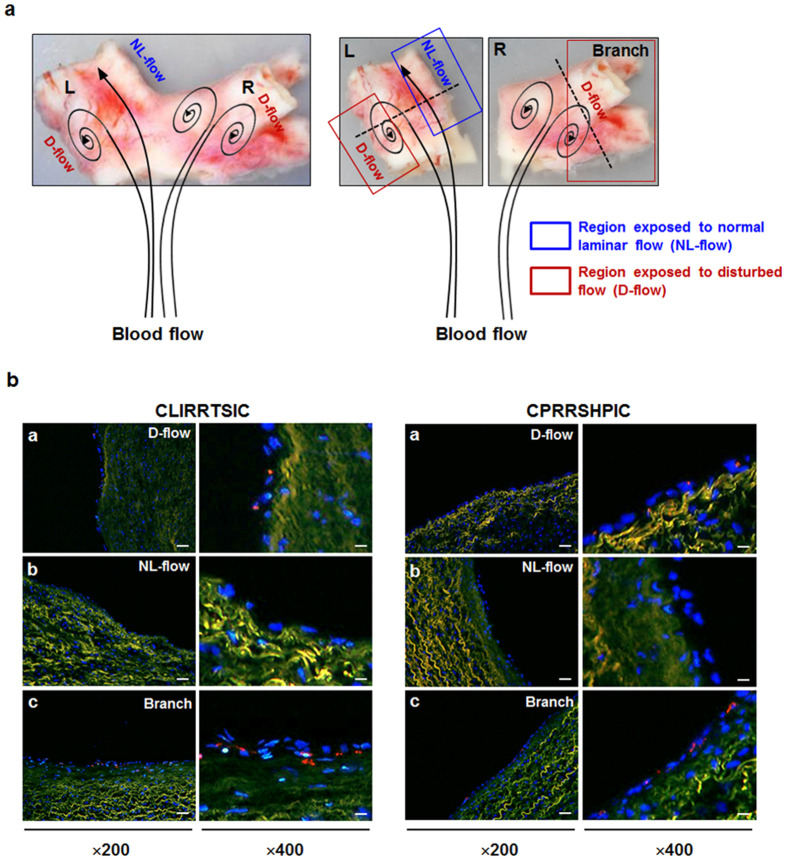
Figure 4. Binding of SPs-displaying phages in human tissue.
Frozen sections of human tissues were incubated with 2 × 10¹¹ pfu of CLIRRTSIC- or CPRRSHPIC-displaying phages for 3 h. (a) Appearance of an isolated human pulmonary artery. Human pulmonary artery was divided into two parts: the straight left (L) and right (R) parts with branch. The black arrow diagram appears the direction of blood flow. (b) Immunofluorescence staining for detection of phage binding in human tissue. Phage binding was compared in three regions with different wall shear stresses: the outer side with disturbed flow and inner side with normal laminar flow of the bifurcation site in left part (L) of specimen, and the branching regions with disturbed flow (Branch) in right part (R) of specimen using confocal microscopy (red: phage, blue: nuclei, green: elastic lamina of vessel) (magnification, ×200; scale bars, 20 μm, ×400; scale bars, 10 μm). Representative images are shown.

