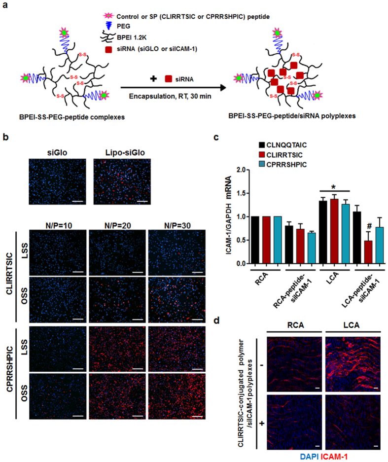
Figure 5. In vitro and in vivo delivery of siRNA to pro-atherogenic ECs using SPs-conjugated polymers.
(a) Schematic diagram of BPEI-SS-PEG-SPs/siRNA polyplexes preparation. The SPs-conjugated polymers with fluorophore-labeled siRNA or anti-intercellular adhesion molecule-1 siRNA (siICAM-1) were prepared by mixing the components for 30 min at room temperature. (b) In vitro delivery of siRNA using peptides-conjugated polymers. HUVECs were exposed to static or flow conditions for 48 h. Cells were incubated with CLIRRTSIC- or CPRRSHPIC-conjugated polymer/siGLO polyplexes for 4 h at various N/P ratios. After 24 h, internalization of siGLO (red) was detected by immunofluorescence microscopy. Nuclei were stained with DAPI (blue) (magnification, ×100; scale bars, 100 μm). Representative images from at least three experiments are shown. (c,d) In vivo delivery of siRNA using peptides-conjugated polymers. siICAM-1 was encapsulated using CLIRRTSIC-, CPRRSHPIC- or control peptide CLNQQTAIC-conjugated polymers. Each peptide-conjugated polymer/siICAM-1 polyplexes were injected intravenously into mice (n = 5) at 3 days after partial ligation of the carotid artery. Twenty-four hours (c) after injection, mice were sacrificed and endothelial enriched RNAs obtained from LCA and RCA. The ICAM-1 mRNA level was determined by qPCR. Shown are means ± SEM of three independent experiments. *, #Significant difference (*RCA vs. LCA; #LCA vs. LCA-peptide-siICAM-1, p < 0.05). Fourty-eight hours (d) after injection with CLIRRTSIC-conjugated polymer/siICAM-1 polyplexes, the carotid arteries were isolated and the protein levels of ICAM-1 evaluated by en face staining. Carotid arteries were fixed and ICAM-1 was stained using an anti-ICAM-1 antibody (red); nuclei were stained with DAPI (blue). Expression of ICAM-1 was observed by confocal microscopy (magnification, ×400; scale bars, 10 μm). Representative images are shown.

