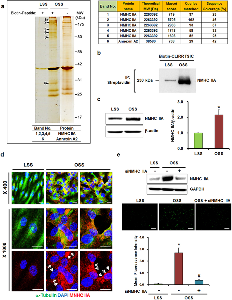
Figure 6. Identification of CLIRRTSIC peptide-binding target proteins.
HUVECs were exposed to LSS or OSS for 48 h, and then incubated with or without biotinylated CLIRRTSIC peptide (20 μM) for 4 h. Cell lysates were incubated with streptavidin beads to pull down the biotinylated CLIRRTSIC peptide-binding proteins and were resolved by SDS-PAGE. (a) Proteomic analysis of CLIRRTSIC peptide-binding target protein. The gel was silver-stained and showed. Six proteins that were selected for sequencing by liquid chromatography-tandem mass spectrometry. MS/MS spectra of [M+H]+ ions of one of the peptides derived from the indicated protein is shown. (b) Binding of CLIRRTSIC peptide to NMHC IIA. HUVECs exposed to LSS or OSS for 48 h were incubated with biotinylated-CLIRRTSIC peptide for 4 h. The immunoprecipitates with streptavidin-beads were immunoblotted using an anti-NMHC IIA antibody. Shown is a representative of three independent experiments. (c,d) Expression of NMHC IIA in ECs. HUVECs exposed to LSS or OSS for 48 h. Cell lysates were immunoblotted and using an anti-NMHC IIA antibody (c). *Significant difference (LSS vs. OSS, p < 0.05). HUVECs exposed to LSS or OSS for 48 h were stained with anti-alpha tubulin and anti-NMHC IIA antibodies and observed using confocal microscopy (d). Shown are representative images of at least three experiments. Anti-alpha tubulin (green), anti-NMHC IIA antibody (red, arrows) and DAPI (blue) (magnification, ×400 or ×1000; scale bars, 10 μm). (e) HUVECs were transfected with siRNA of NMHC IIA (50 nM) for 48 h. After transfection, Cells were exposed to LSS or OSS for 48 h and then incubated with biotin-labeled CLIRRTSIC peptide (20 μM) for 4 h. Attached peptides were stained with streptavidin conjugated Qdot (green) (magnification, ×100; scale bars, 100 μm). Representative images are shown. Relative mean fluorescence intensity calculated using Image J for binding of CLIRRTSIC peptide (green). *, #Significant difference (*LSS vs. OSS; #OSS vs. OSS + siNMHC IIA, p < 0.05).

