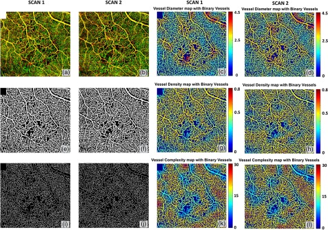
Fig. 6.
Quantitative OMAG analysis of a BRVO case with repeated scans. (a) and (d) OMAG image of occluded region, covering a FOV of . (b) and (e) Vessel area map. (c) and (f) Vessel skeleton map. (g) and (j) Vessel density map integrated with vessel area map. (h) and (k) Vessel diameter map integrated with vessel area map. (i) and (l) Vessel complexity map integrated with vessel area map.

