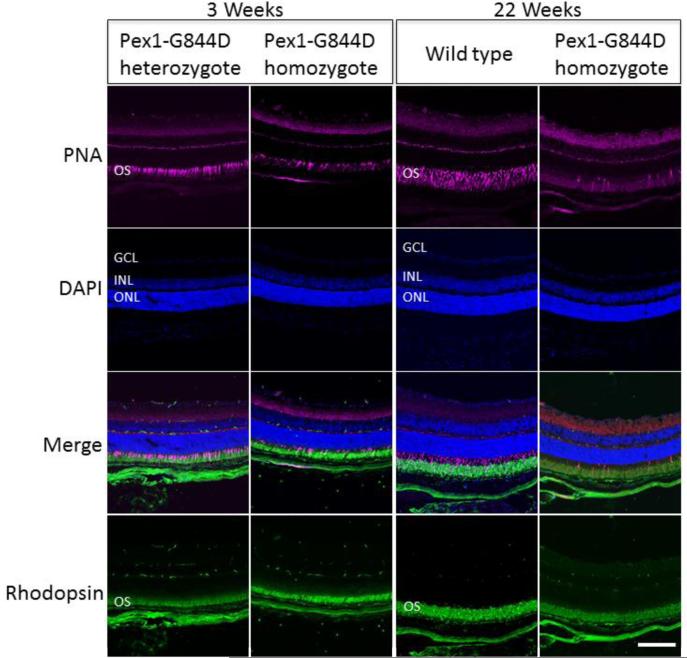
Figure 5.
Retinal Histology. Immunostaining of photoreceptor outer segments was performed for the Pex1-G884D homozygote and control mouse retinas at three-weeks and 22-weeks. Peanut agglutinin (PNA; magenta) and an antibody against rhodopsin (green) were used to stain cone and rod outer segments, respectively. Nuclei were counter-stained with DAPI (blue). Retinal layers are marked by these abbreviations: GCL, ganglion cells layer; INL, inner nuclear layer; ONL, outer nuclear layer; and, OS, outer segment. Scale bar equals100 mm. Immunostaining with the cone specific PNA in 3-weeks-old mice shows that some cone photoreceptors are preserved in Pex1-G844D homozygotes. In contrast, at 22-weeks of age PNA staining in the Pex1-G844D homozygotes is consistent with a loss of cone photoreceptors. The rhodopsin staining was not as robust in the Pex1-G844D homozygotes as the control indicating that the retinal degeneration may not be limited to the cone photoreceptors.

