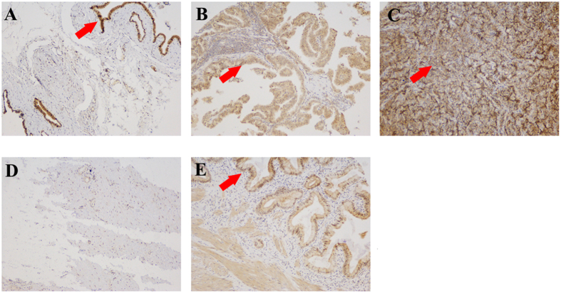
Figure 1. PKM2 expression in sections of cancerous gallbladder tissue.
(A) Well-differentiated adenocarcinoma exhibited low PKM2 staining and was given a score of 2. (B) Moderately differentiated adenocarcinoma exhibited moderate PKM2 expression and was given a score of 3. (C) Poorly differentiated adenocarcinoma exhibited strong PKM2 expression and was given a score of 4. (D) Gallbladder tissue was used as the negative control as it exhibited negative staining for PKM2 and was given a score of 0. (E) Gallbladder tissue which were positive staining for PKM2 and was given a score of 1 (F) Western blot with an antiPKM2 antibody revealed positive expression of PKM2 in GBC-SD, NOZ and SGC-996 cells. The red arrow in (A–C,E) point to the positive stained cell which were dyed brown. Original magnification: ×200.

