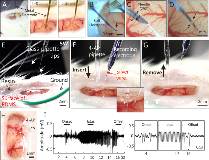
Figure 2. Accessing neural tissue directly through the PDMS cranial window over a rat brain.
(A) A side view of the metal electrode penetration directly through the PDMS and its magnified images at time series. The injected electrode was slowly pulled out at a speed of 0.5 mm/min. No bleeding or CSF leakage was observed. (B–D) A soft and thin type of the multi-channel electrode alone could not penetrate (B) and bent upon contact with the PDMS surface (black arrow). (C) Penetration was aided by a sharp needle (29 gauge) and (D) the electrode was slid inside via a slit (black arrows). (E–H) Sequential images of the glass pipette placement and removal performed at 5 weeks post-cranial window implantation. Two glass pipettes were inserted into the brain tissue directly through the PDMS cover by avoiding major vasculature; one glass pipette (clear) was used to inject 700 nl of potassium blocker 4-aminopyridine (4-AP) at 15 mM concentration, to induce seizure activities, while the other pipette (blue with trypan blue dye for visualization) contained a silver wire filled with 0.9% NaCl, which was used to record local field potential (LFP). Tips of both pipettes were marked with arrowheads in E and outlined with dashed lines in an inserted, expanded image of (F). No noticeable leakage was detected after the pipettes were removed from the cranial window (G,H). To prevent the possible rupture of PDMS later, the insertion sites were glued with fast-acting adhesive cyanoacrylate (H). (I) Record electrophysiological signals induced by 4-AP. We observed a clear seizure onset, ictus, and offset. Expanded time courses shown at bottom right were obtained from three line-marked time periods.

