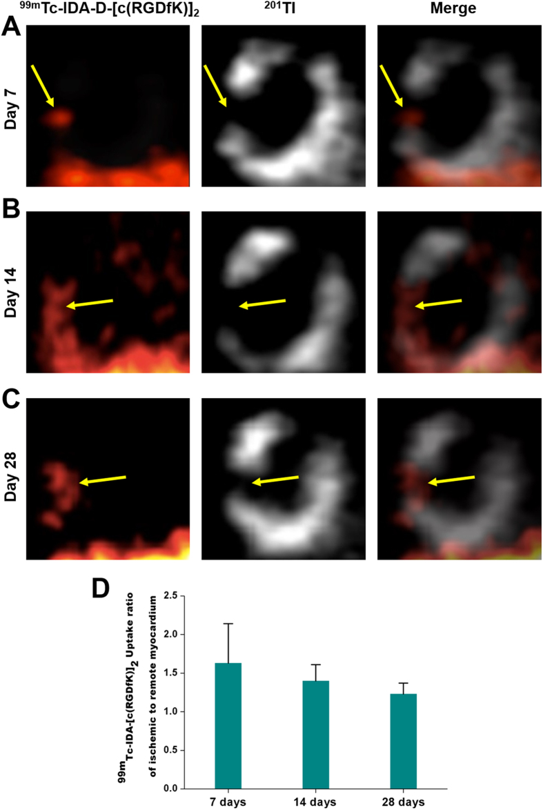
Figure 2. In vivo SPECT imaging of MI/reperfusion model with 201Tl and 99mTc-IDA-D-[c(RGDfK)]2.
(A–C) Serial SPECT scans were acquired at 7 (A), 14 (B), and 28 (C) days after MI/reperfusion. Shown are representative vertical long axis images from four independent experiments (n = 4 rats). The transiently ischemic, but still viable myocardium (arrows) was noted by strong signal of 99mTc-IDA-D-[c(RGDfK)]2 (left column) and defect of perfusion signal, 201Tl (middle column). Images from two energy windows (99mTc-IDA-D-[c(RGDfK)]2: 130–150 keV, 201Tl: 60–90 keV) were merged (right column) to identify localization of dual isotopes. (D) 99mTc-IDA-D-[c(RGDfK)]2 uptake ratio of ischemic to remote myocardium was peak at day 7 (1.63 ± 0.51) and gradually decreased, but high enough for clear differentiation even 28 days later. Data are means ± SD (n = 4).

