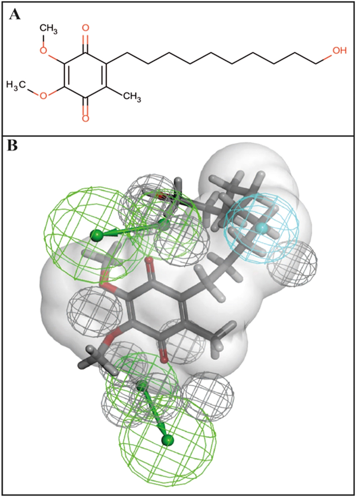
Figure 4. Idebenone mapped to the ThyX N = 18 pharmacophore model.

(A) idebenone 2D structure. (B) Pharmacophore showing van der Waals surface based on C8-C1, Blue = hydrophobe, green = hydrogen bond acceptor, grey = excluded volume.

(A) idebenone 2D structure. (B) Pharmacophore showing van der Waals surface based on C8-C1, Blue = hydrophobe, green = hydrogen bond acceptor, grey = excluded volume.