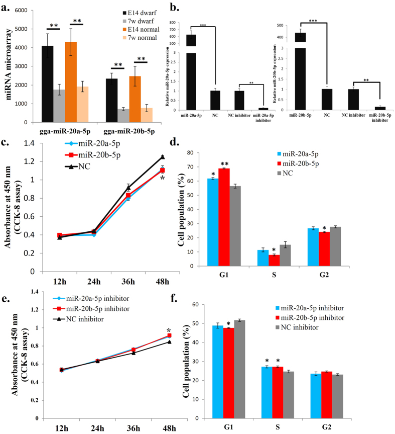
Figure 1. miR-20a-5p and miR-20b-5p repress myoblast proliferation.
(a) The miRNA microarray hybridization signals of miR-20a-5p and miR-20b-5p in E14 and 7w of dwarf and normal chickens’ leg muscle. (b) The expression of miR-20a-5p and miR-20b-5p after transfection of the indicated miRNA mimic or miRNA inhibitor. (c) CCK-8 assay was performed to access the effect of miR-20a-5p and miR-20b-5p overexpression on myoblast proliferation. (d) Cell cycle analysis of myoblasts at 48 h after transfection of miR-20a-5p and miR-20b-5p mimics or NC mimic. (e) CCK-8 assay was performed to access the effect of miR-20a-5p and miR-20b-5p loss-of-function on myoblast proliferation. (f) Cell cycle analysis of myoblasts at 48 h after transfection of miR-20a-5p inhibitor, miR-20b-5p inhibitor or NC inhibitor. Results are shown as the mean ± sem of three independent experiments. One sample t test was used to analysis the statistical differences between groups. *p < 0.05; **p < 0.01; ***p < 0.001.

