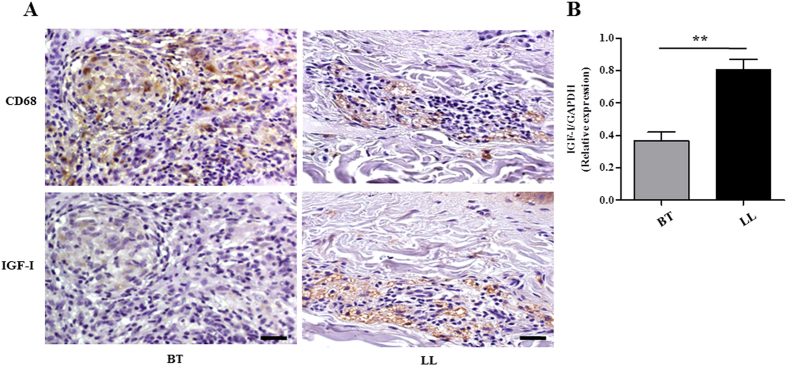
Figure 1. IGF-I is highly expressed in dermal lesions of lepromatous patients.
(A) IGF-I and CD68 expressions were evaluated in serial sections of borderline (BT) and lepromatous (LL) skin lesions by immunohistochemical staining with diaminobenzidine. Immunolabelled cells appear as brown stained. Hematoxylin counterstained nuclei appear in blue. Data shown are representative of three lesions of each clinical form evaluated. Scale bar = 20 μm. (B) Comparative analysis of IGF-I mRNA expression in skin biopsies of BT (n = 7) and LL (n = 9) lesions by qRT-PCR. Data are shown as mean ± SE. Student’s t-test was performed and used for statistical analysis. **p < 0.005.

