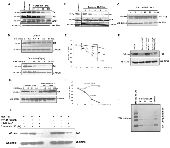
Figure 1. Curcumin decreased HIV-1 Tat protein.
(A) HEK-293T cells were transfected with 1 μg of Myc-Tat expressing plasmid, and after 36 hrs treated with curcumin for 8 hrs, lysed and probed for Tat, p53 and GAPDH. pEGFP-N1 (50 ng) was also transfected as transfection control. The blot shown is a representative of three independent experiments.(B) Myc-Tat was transfected in HEK-293T cells and curcumin treatment was performed for increasing time period. The blot is a representative of three independent experiments. (C) Gag-Opt (1 μg) was transfected and curcumin treatment was performed followed by immmuno-blotting for Gag protein in HHEK-293T cells. (D) Myc-Tat transfected HEK-293T cells were treated with CHX alone or with curcumin for time periods as indicated and Tat protein level was measured by western blotting. (E) The mean value of Tat protein from three independent experiments was plotted with respect to treatment period. P value was calculated by a two-tailed t-test (*P < 0.05, **P < 0.01; NS, not significant). (F) The Myc-Tat transfected HEK-293T cells were treated with curcumin, along with proteasomal and lysosomal inhibitors MG132 and ammonium chloride for 8 hrs, subsequently Tat level was measured. (G) Myc-Tat transfected HEK-293T cells were treated with curcumin and CHX in the absence or presence of MG132 for different time periods followed by western blotting for Tat protein. (H) Densitometry of Tat bands was carried out by using image J and plotted with respect to treatment period. (I) HEK-293T cells were transfected with 1 μg of Myc-Tat (lanes 1–3) for 36 hrs followed by treatment with curcumin and Pyr-41 for 6 hrs subsequently the western blotting was done for Tat protein. HEK-293T cells were transfected with 1 μg of Myc-Tat and 2 μg of HA-Ub KO, 36 hrs of transfection curcumin treatment was done for 6 hrs and Tat protein was blotted. (J) HEK-293T cells were transfected with 1 μg of Myc-Tat and 2 μg of 6X-His Ubiquitin plasmid, after 36 hrs the cells were treated with increasing dose of curcumin or MG132. The ubiquitinated proteins were purified using Ni-NTA affinity chromatography and ubiquitinated Tat was blotted using anti-Myc antibody. The original uncropped blot images for (A–D,F,G,I,J) are shown in supplementary information as supplementary figure.

