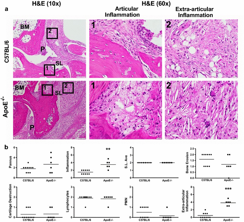
Fig. 2.

Increased articular and extra-articular inflammation is present in ApoE−/− mice. a H&E staining of arthritic ankle joints from C57BL/6 (control, n = 10) and ApoE−/− (n = 6) mice. Boxes 1 and 2 demarcate articular and extra-articular areas of high magnification (×60), respectively. b Histopathological scores for pannus formation, inflammation, synovial lining average, bone erosion, cartilage destruction, lymphocytes, polymorphonuclear cells and extra-articular inflammation on ankle joints from above. Data are represented as mean ± SEM. Asterisk denotes statistically significant differences: **p < 0.01, ***p < 0.001. BM bone marrow, SL synovial lining, P pannus
