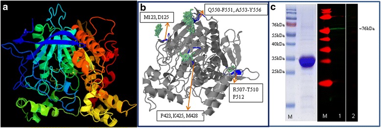
Fig. 3.

a Three dimensional model of S. japonicum acetylcholinesterase determined using PHYRE2. Image coloured by rainbow from N to C terminus, Model dimensions (Å) X: 61.705 Y: 62.361 Z: 71.856 are the same as that of ShAChE. b The predicted binding sites of SjAChE with N-Acetylglucosamine (NAG). The four predicted N-Acetylglucosamine binding sites (in blue) are located at (i) M123, D125; (ii) P423, K245, M428; (iii) R507-T510, P512; and (iv) Q550-F551, A553-Y556 in SjAChE. The NAG residues are shown in green. c Western blot analysis using anti- SjAChEC to detect the total extracts from adult S. japonicum. Left panel: SDS-PAGE gel of purified recombinant protein SjAChEC (Molecular size: 30 Kda); Right panel: western blot analysis of total extract from adult S. japonicum worms. The protein extract was probed with rabbit anti-SjAChEC antibody (Lane 1) by recognising a band of approximately 76 kDa which match the calculated molecular size for native SjAChE Pre-immune sera (Lane 2) was used as control. Lane M, PageRulerTM pre-stained protein ladder
