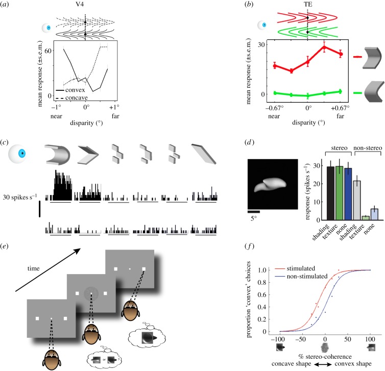
Figure 3.
3D-shape representations in the anterior ventral pathway. (a) Average response of a V4 neuron to convex (solid line) and concave (dashed line) stimuli at different positions in depth relative to the fixation plane (x-axis; see pictograms above). This neuron changed its stimulus preference depending on the position-in-depth of the stimulus. Its tuning curve was similar to that obtained when a flat stimulus was varied across positions in depth (not shown), indicating that 3D shape was not driving this neuron's response. (Figure adapted with permission from Hegdé & Van Essen [63].) (b) Average response of a TE neuron to concave stimuli (top row; red) or convex stimuli (bottom row; green) placed at different positions in depth relative to the fixation plane (dotted line). Left: in front of the fixation plane. Right: behind the fixation plane. This neuron retained its concave-shape preference across different positions in depth. (Figure adapted with permission from Orban et al. [11].) (c) Average response of a TE neuron to concave (top row) and convex (bottom row) stimuli. The different columns indicate different approximations to the smoothly curved stimulus on the left (see pictograms above). This neuron preferred smoothly-curved concave 3D shapes. (Figure adapted with permission from Janssen et al. [64].) (d) Yamane et al. [65] used a genetic algorithm to optimize stimuli to individual TE neurons. They found that neurons in TE prefer fairly complex 3D shapes (see example on left). The labels below or above the bar plots show which depth cues (disparity, shading or texture) were present in the stimulus. Removing disparity significantly reduced this neuron's response (compare left three and right three bars), illustrating the importance of depth for shape representations in TE. (Figure adapted with permission from [65].) (e) 3D-shape categorization task. See figure 1c for an example stimulus. Monkeys were trained to categorize disparity-defined 3D shapes as either convex or concave by making an eye movement to the left (convex) or right (concave) response target to indicate the perceived shape. (f) Example microstimulation session in TE. Proportion ‘convex’ choices is plotted as a function of the stimulus (right x-axis: convex shape, left: concave shape) and the noise applied to the stimulus (% stereo-coherence). Blue colour shows performance on trials without stimulation, red colour on trials with stimulation. Stimulation was applied on 50% of randomly chosen trials, and only during stimulus presentation. When clusters of TE neurons with a convex 3D-shape preference were electrically stimulated while the monkey performed the 3D-shape categorization task, the monkey more often reported perceiving a convex shape. (Figure adapted with permission from Verhoef et al. [66].)

