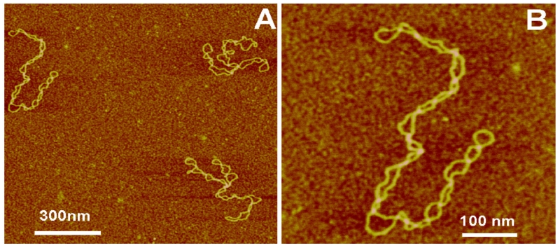
FIG. 1.
AFM images of supercoiled DNA prepared using AP mica. (A) A large-scale image with three DNA molecules in the field. (B) Zoomed image of the top-left molecule in (A). The supercoiled molecules appear as uniformly twisted filaments with a plectonemic shape [reprinted with permission from Elsevier, Copyright 2011].7

