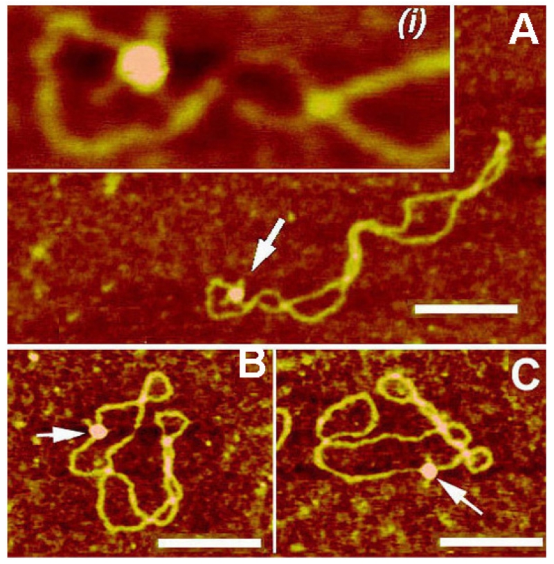
FIG. 3.
AFM images of cruciforms in their complexes with RuvA protein. (A) pUC8F14C plasmid DNA. Cruciforms are indicated with arrows and numbered. (B–D) pUC8F14C plasmid DNA with RuvA protein. Cruciforms in complexes with the protein are indicated with arrows. The scale bar is 100 nm. [reprinted with permission from Elsevier, Copyright 2000].36

