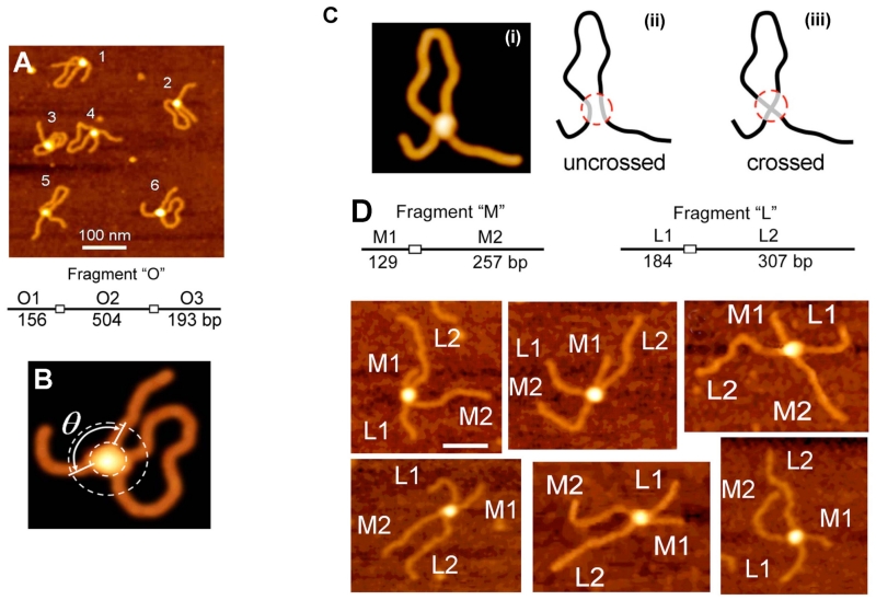
FIG. 5.
AFM study of SfiI-DNA complexes. (A) and (B) AFM images of synaptic SfiI-DNA complexes formed between two recognition sites separated by 504 bp (left). (C) Models of the possible arrangement of DNA strands in the synaptic complex SfiI-DNA. (D) Complexes formed by SfiI by synapsis of M and L DNA fragments (trans type complexes). The schematic for the fragments is shown on the top and the AFM images are below. The flanks M1, M2 and L1, L2 are assigned according to their length measurements [reprinted with permission from the American Chemical Society, Copyright 2006].54

