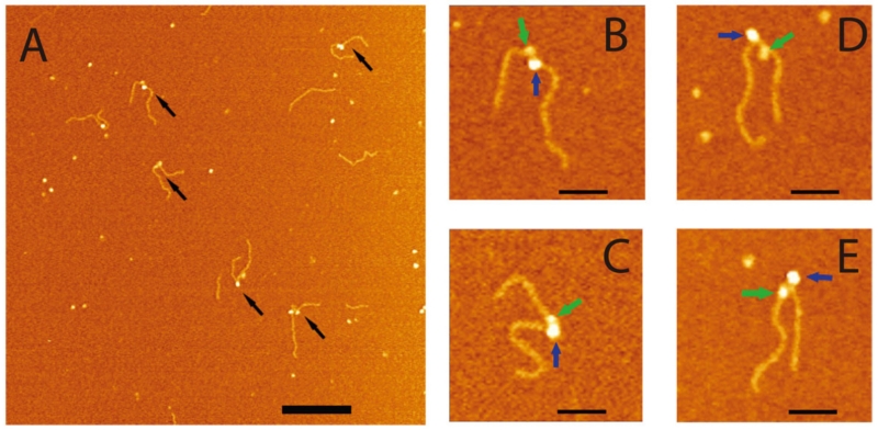
FIG. 7.
AFM images of the complexes made by SSB and RecG with the fork DNA substrate. (A) Large scale AFM images, in which double-particle features are indicated with arrows. Bar size is 200 nm. Zoomed images (B-E; bar size = 50 nm) of four double-particle complexes. Large and small particles are indicated with blue and green arrows, respectively. The black and green arrows point to SSB and RecG proteins in the double-particle complexes. The figure was reproduced from paper respectively [reprinted with permission form Nature Publishing Group, Macmillan Publishers Ltd. Creative commons license, Copyright 2015].62

