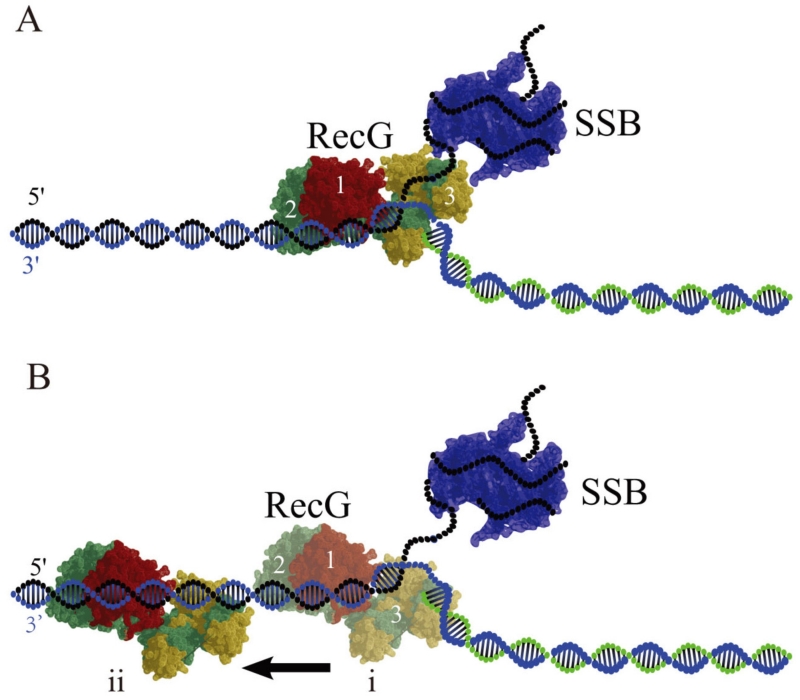
FIG. 8.
Models for RecG binding to the DNA fork in the presence of SSB. (A) No interaction with SSB. In the type 1 complex, RecG binds specifically to all arms of the fork, with domains 1 and 2 binding the parental DNA duplex, whereas the wedge domain 3 binds the single-stranded arm of the fork. SSB occupies the ssDNA arm of the fork. (B) In the type 2 complex formed after SSB remodeling, the wedge domain 3 dissociates from the ssDNA arm, but domains 1 and 2 remain bound to the parental DNA flank, enabling RecG to translocate along the duplex as shown in (i) and (ii). Model (i) shows the RecG position at the fork just after remodeling and (ii) shows the RecG position after translocation away from the fork respectively [reprinted with permission form Nature Publishing Group, Macmillan Publishers, Ltd. Creative commons license, Copyright 2015].62

