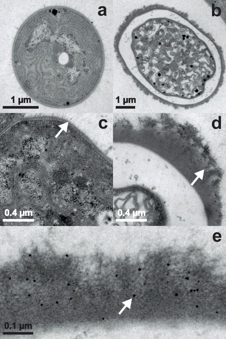
Figure 3.
TEM images of Anabaena cylindrica incubated with an overall concentration of 0.8 mM Au3+ for 15 minutes. Vegetative cells are displayed in panels (a) and (c), heterocysts in panels (b), (d) and (e). The white arrows guide the eye to one already formed nanoparticle in each panel with larger magnification.

