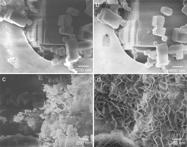
Figure 5.
Helium ion microscopy images of vapor-phase-grown MgO cubes before (a,c) and after (b,d) exposure to liquid water. The sample areas imaged in (a) and (c) are the same as the ones imaged in (b) and (d), respectively. Clearly, different types of nanostructures can result from the interaction of MgO particle surfaces with H2O depending on their size and distribution at the sample surface. Comparison between (a) and (b) points to the volume expansion during hydration and hydroxylation, while (c) and (d) show a probable dissolution/precipitation mechanism. (HIM SE images recorded at 30 kV acceleration voltage and (a,c) 0.3 pA beam current, 1.4·1015 cm−2 ion dose, (b,d) 0.1 pA beam current, 1.2·1015 cm−2 ion dose.)

