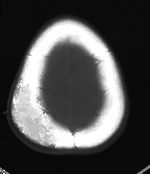
Figure 3.

Noncontrast computed tomography head (axial view and bone window) image showing the hypodense lesions with sharp, thickened, sclerotic margins

Noncontrast computed tomography head (axial view and bone window) image showing the hypodense lesions with sharp, thickened, sclerotic margins