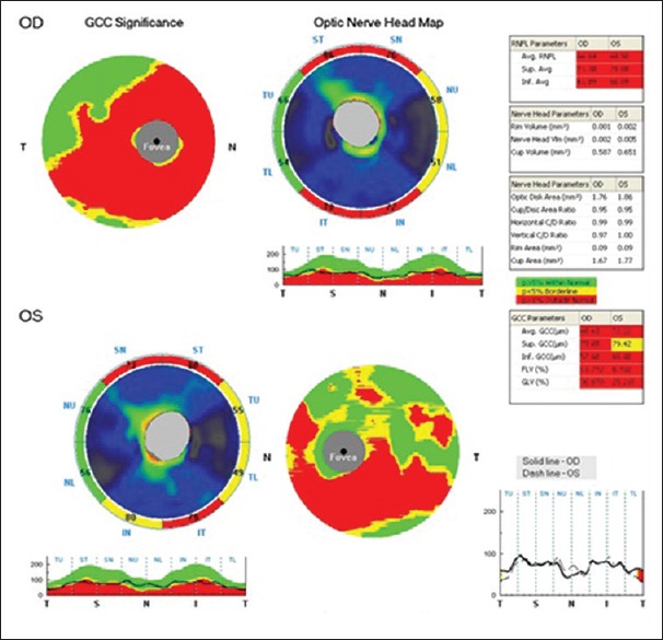
Figure 1.

Optical coherence tomography pictures of prototype patient. Optical coherence tomography pictures showing normative values of five parameters of ganglion cell complex and three parameters of retinal nerve fiber layer. This picture shows thinning of superior and inferior retinal nerve fiber layer and corresponding thinning of ganglion cell complex
