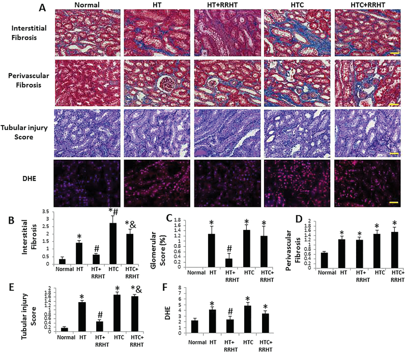
Figure 2.
Renal tissue remodeling. (A) Representative renal trichrome, periodic acid Schiff (PAS), and dihydroethidium (DHE) staining (all ×20). Renal interstitial fibrosis, glomerulosclerosis, and tubular injury increased in renovascular hypertension (HT), and HT and hypercholesterolemia (HTC) kidneys. After reversal of renovascular HT (RRHT), interstitial fibrosis and tubular injury decreased in HT, but not in HTC+RRHT (B, C, and E). Perivascular fibrosis was similarly increased in the HT, HT+RRHT, HTC, and HTC+RRHT groups (D). DHE staining was elevated in HT, HTC, and HTC+RRHT, suggesting increased oxidative stress, which decreased in HT+RRHT (F). *P < 0.05 vs. Normal; # P < 0.05 vs. HT; & P < 0.05 vs. HT+RRHT. Scale bar = 50 µm.

