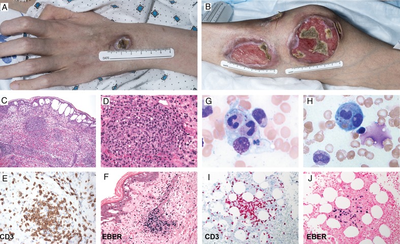
Figure 1.
A, B, Skin lesions in patient 5 due to Epstein-Barr virus (EBV)–positive hydroa vacciniforme–like lymphoma. C–F, Hematoxylin-eosin staining show fluid-filled vacuoles in the epidermis (C) and a dense lymphocytic infiltrate (D), CD3 T cells (E), and EBV-encoded RNA (EBER)–positive cells in the skin (F). G, H, Hemophagocytosis. I, CD3 T cells. J, EBER-positive cells in the bone marrow.

