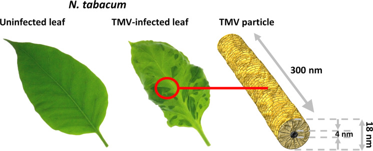
Figure 1.
Tobacco mosaic virus infection. Left: Leaf of a healthy tobacco plant (Nicotiana tabacum 'Samsun' nn). Center: Leaf of a TMV-infected plant showing the typical TMV-associated mosaic (light and dark green mottling on the leaf blade). Right: Organization and dimensions of a TMV ribonucleoprotein particle, with the RNA not depicted as it is completely enclosed in the CP helix (golden).

