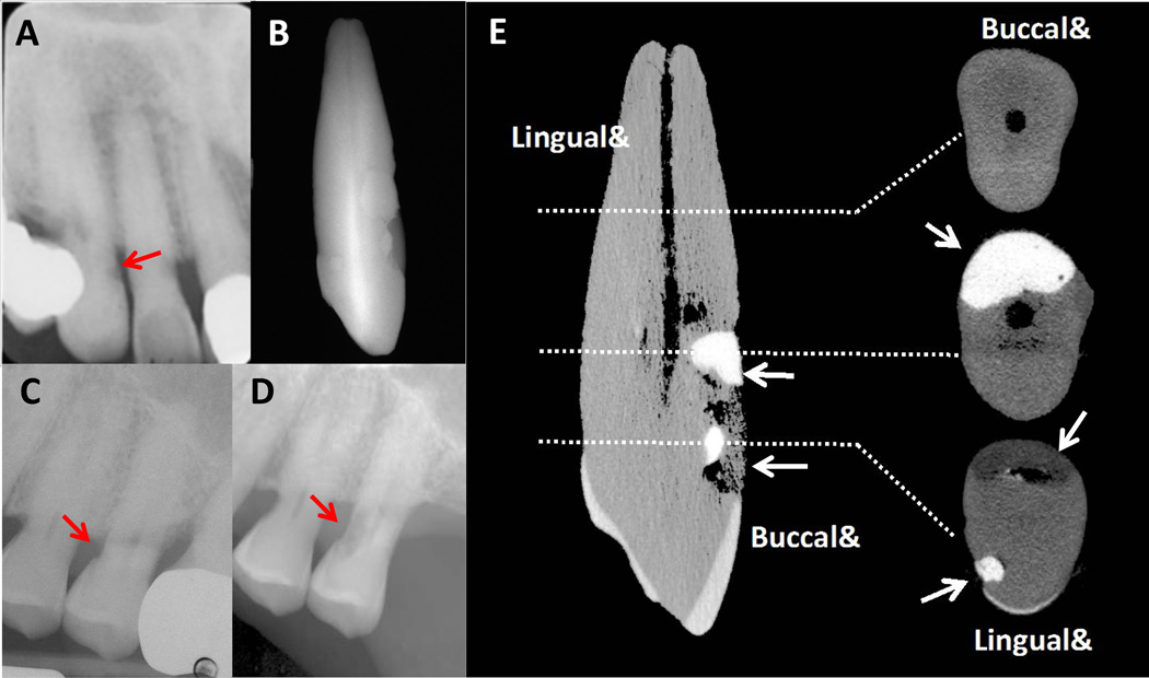
Figure 3.
A. Radiograph of tooth #6 from the proband showing cervical root resorption (arrow). B. Radiograph of tooth #6 after extraction (2006). Note the presence of multiple resin restorations on the buccal and palatal surfaces. C. Radiograph of tooth #13 from the proband (2005). Resorption (arrow) is present on the palatal and mesial aspect of the tooth. D. Radiograph of tooth #13 showing progression of resorptive lesion (arrow) on the mesial surface just prior to extraction (2008). E. Micro-CT image of tooth #6 from the proband. The image on the left shows the entire tooth. Images on the right represent cross-sectional slices of the tooth. Despite the invasive nature of the resorption, there is no perforation into the pulp chamber. Arrows point towards resin restorations. Note: 3A adapted from “A Familial Pattern of Multiple Idiopathic Cervical Root Resorption in a Father and Son: A 22-Year Follow-Up”.21 Adapted with permission from the American Academy of Periodontology.

