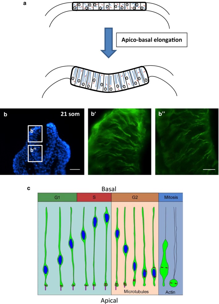
Figure 2.

Role of MTs in apico‐basal elongation and INM, in the vertebrate neural plate. (a) Paraxial alignment of MTs causes epiblast cells to undergo apico‐basal elongation, producing the high columnar shape that characterizes cells of the early bending neural plate in birds, amphibia and mammals. Images are schematic transverse sections. (b) Immunohistochemistry (green) for α‐tubulin in transverse sections of the closing mouse spinal neural plate at the 21 somite stage. Apico‐basally aligned MTs are visible in both the dorsal (b') and ventral (b'') regions. Blue: DAPI‐stained nuclei. Scale bars: 80 μm (b); 20 μm (b', b''). (c) During INM, nuclei (blue) move basally during the G1‐phase and remain at the basal neuroepithelial surface during the S‐phase. During the S‐to‐G2 transition, dynein is activated and moves the nucleus toward the minus end of MTs at the centrosome (red), which is rooted in an apical cilium (pink). During the G2‐phase, the cilium disassembles, allowing the newly‐untethered centrosome to relocate to the nucleus, where it initiates mitosis. (c) Reproduced with permission from fig. 6 of Spear & Erickson, 2012, Developmental Biology 370: 33–41.
