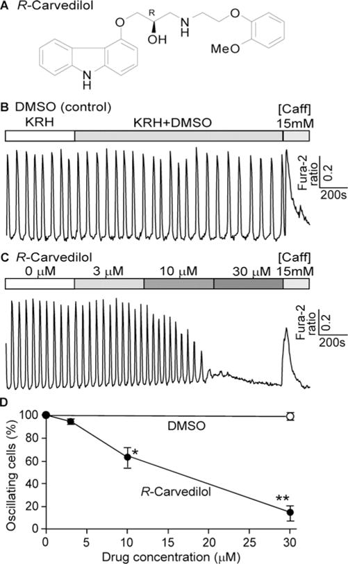
Figure 1. R-carvedilol inhibits spontaneous Ca2+ release in HEK293 cells.

(A) Chemical structure of R-carvedilol. (B and C) Stable inducible HEK293 cells expressing the RyR2-R4496C mutant were loaded with 5 μM fura2/AM in KRH buffer. Representative traces of fura 2 ratios in HEK293 cells (∼197–419) perfused with 1 mM extracellular Ca2+ in KRH buffer containing DMSO (B) or various concentrations of R-carvedilol (0, 3, 10 and 30 μM) are shown. (C) Percentage of cells showing spontaneous Ca2+ oscillations in cells treated with DMSO (control) or R-carvedilol. Data shown are means ± S.E.M. (n=5–7; * P < 0.05, **P < 0.001 compared with DMSO).
