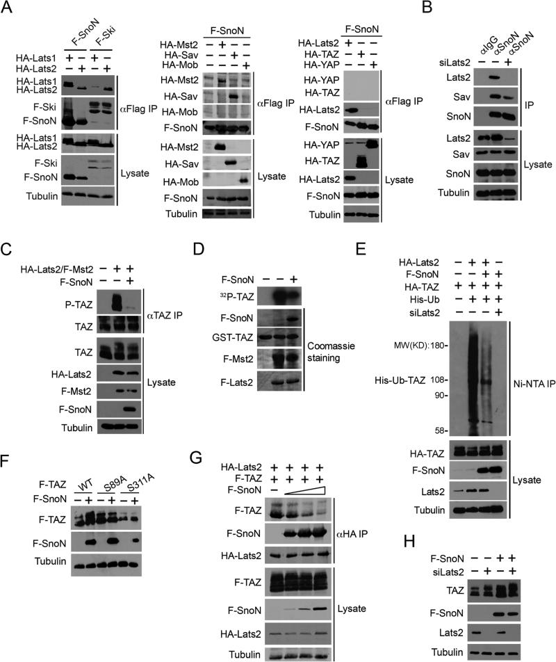
Figure 5. SnoN interacts with the Hippo kinase complex and inhibits the ability of Lats2 to phosphorylate TAZ.
(A) F-SnoN or F-Ski was isolated by immunoprecipitation (IP) with anti-Flag from 293T cells transfected with F-SnoN or F-Ski together with Hippo signaling molecules as indicated, and proteins associated with F-SnoN or F-Ski were detected by Western blotting with anti-HA antibodies (upper panels). The abundance of these proteins in the lysates was shown in the lower panels. Tubulin was used as a loading control. (B) MCF10A cells were transfected with either scramble control or siLats2 and treated with 20 μM MG132 for an additional 6 h. Interactions between endogenous SnoN and Hippo signaling proteins were examined by IP with anti-SnoN or anti-IgG followed by Western blotting with antibodies specific for individual Hippo components (upper). The abundance of these proteins in the cell lysates was assessed by Western blotting (lower). (C) HA-Lats2 and F-Mst2 were transfected into MCF10A cells in the absence or presence of F-SnoN and treated with 20 μM MG132 for 6 h. Phosphorylated TAZ was isolated by IP with anti-TAZ followed by Western blotting with anti-Phospho-S89 TAZ. (D) In vitro kinase assay. Lats2 and Mst2 were IP from cells transfected with F-Lats2 and F-Mst2 either alone or together with F-SnoN and subjected to in vitro kinase assay using GST-TAZ as a substrate. 32P-TAZ was detected by auto-radiography (top panel). The abundance of GST-TAZ, F-Lats2, F-Mst2 and F-SnoN was measured by Coomassie blue staining (lower panels). (E) 293T cells transfected with various constructs indicated were treated with 20 μM MG132 for 6 h, and His-Ub-TAZ was pulled down by the Ni-NTA column and detected by Western blotting with anti-HA (top panel). The abundance of proteins in the cell lysates was assessed by Western blotting (lower panels). (F) WT, S89A or S311A TAZ were transfected into 293T cells alone or together with F-SnoN, and the expression levels of these proteins analyzed by Western blotting with anti-Flag. (G) 293T cells were transfected with a fixed amount of F-TAZ and HA-Lats2 and increasing amounts (0, 0.5, 1, 2 μg) of F-SnoN and then treated with MG132 for 6 h. SnoN and TAZ that associated with Lats2 were isolated by IP with anti-HA and detected by Western blotting with anti-Flag. (H) Lats2 siRNAs were transfected into control or F-SnoN-overexpressing MCF10A cells. Endogenous TAZ and Lats2 levels were detected by Western blotting.
See also Figure S4.

