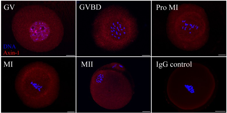
Fig 1. Subcellular localization of Axin-1 during meiotic maturation in mouse oocytes.
Oocytes at various stages were stained with an antibody against Axin-1 (red) and each was counterstained with DAPI to visualize DNA (blue). Key: GV, oocytes at germinal vesicle stage; GVBD, oocytes at germinal vesicle breakdown; Pro MI, oocytes at the first prometaphase stage; MI, oocytes at the first metaphase stage; MII, oocytes at the second metaphase stage. Oocytes in the negative control group were incubated with rabbit IgG. Scale bar = 20 μm.

