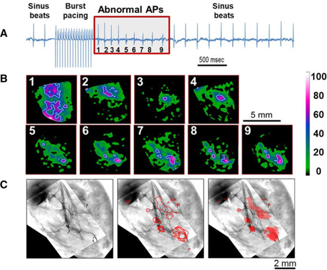
Figure 1.

Subendocardial origins of spontaneous beats. A, Volume-conducted ECG recording of a catecholaminergic polymorphic ventricular tachycardia heart with 3.6 mmol/L Ca2+ and 160 nmol/L isoproterenol before, during, and after 12-Hz burst pacing. Nine abnormal action potentials (APs) were induced after pacing (red box, numbered). B, Earliest right ventricular endocardial AP wave breakthroughs of abnormal beats (1–9, each in a separate box) shown by white and magenta spots (color index bar represents percent from maximal signal in each frame). C, Image of green fluorescent protein–mapped area of the Purkinje network (left) superimposed with white contour lines from B (middle and right). Note colocalization of triggered beats with branches of the Purkinje conduction system.
