
FIGURE 1.
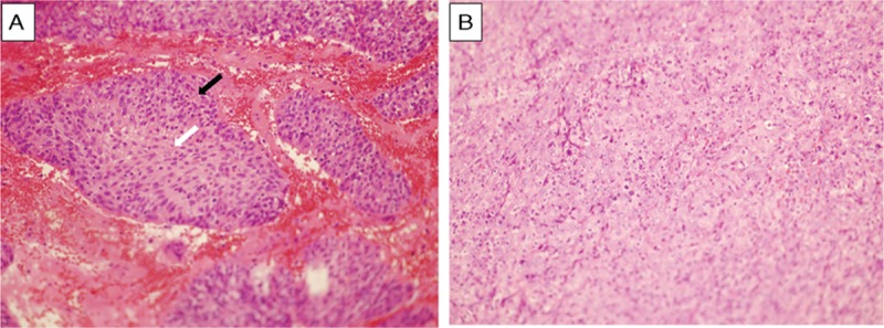
Microscopy morphology of nasopharyngeal nonkeratinizing carcinoma was shown here. (A) Differentiated subtype (40×) showing cellular stratification (black arrow), pavementing, and well-defined cell distinct (white arrow). (B) Undifferentiated subtype (40×) showing syncytial sheets of large tumor cell without distinct border, vesicular nuclei, and large central nucleoli.