
FIGURE 1.
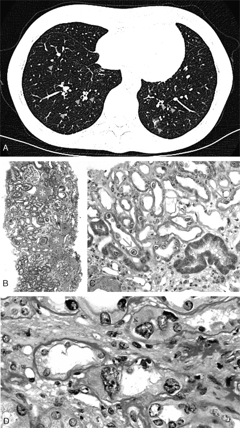
(A) Chest CT-scan showing bilateral bronchectasis with bronchial wall thickening (blue arrowheads). (B) Light microscopy at low magnification using Masson's trichrome staining showed chronic tubulointerstitial nephritis (green asters) with severe fibrosis, tubular atrophy, and inflammatory interstitial infiltrates. Globally sclerotic glomeruli (yellow asters) and severe vascular lesions (red asters) were also observed (original magnification x50). (C) Remarkably, numerous tubular cells in both the cortex and medulla showed nuclear enlargement with irregular outlines (orange arrows, original magnification x200). (D) Periodic acid-Schiff staining showing typical karyomegalic tubular epithelial cells (orange arrows) characterized by markedly enlarged nuclei with irregular outlines, and hyperchromatic and prominent nucleoli (original magnification x600).