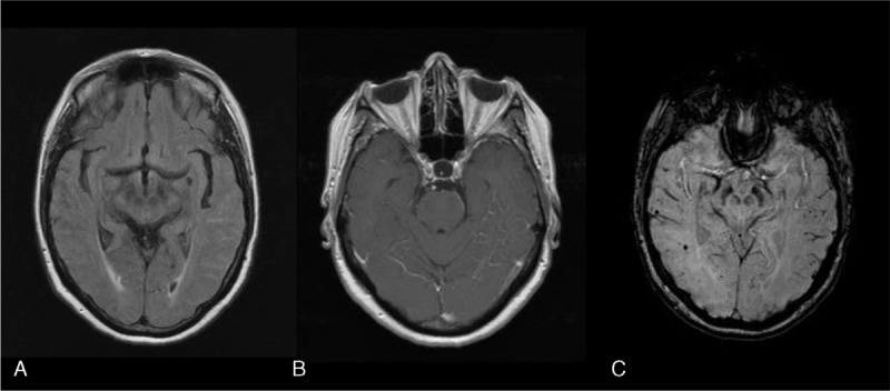
FIGURE 2.

Magnetic resonance imaging of a patient with ABRA showing a strictly leptomeningeal process. A, Fluid Attenuation Inversion Recovery (FLAIR) sequence shows non nulling of the subarachnoid signal involving the left temporal lobe consistent with a leptomeningeal process. No underlying white matter abnormalities. B, Post Gadolinium T1-weighted images shows avid leptomeningeal enhancement. C, Axial susceptibility weighted imaging (SWI) shows multiple bilateral cortical-subcortical microhemorrhages throughout cerebral hemispheres.
