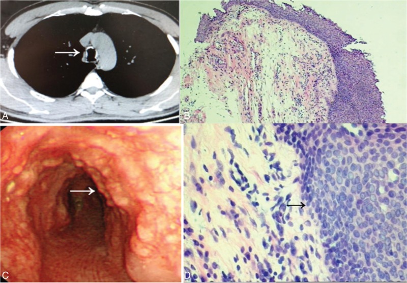
FIGURE 2.

(A) CT scan of case 1 demonstrates irregularly thickened and abroad calcified nodules of the trachea. Arrow shows the calcific ring. (B) Bronchoscopy findings show multiple polypoid, white lesions covered with the anterior and lateral walls of trachea. Arrow shows the calcific nodules. (C and D) Histological appearance indicates infiltration of chronic inflammatory cells and arrow shows diffuse epithelial squamous metaplasia in submucosa (HE staining, original magnification ×100, ×400).
