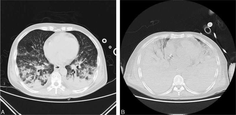
FIGURE 1.

Computed tomography of the chest with contrast. (A) Computed tomography of the chest on day 1 of admission. It revealed widespread bilateral infiltrates surrounded by ground-glass opacification. (B) Computed tomography of the chest on day 4 of admission. It revealed extensive bilateral pulmonary infiltrates, which was worse than the first day.
