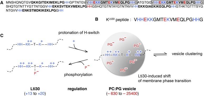
Figure 1.
Schematic image of disordered Lti30 binding to lipid (PC:PG) vesicles. Blue represents a positive charge and red represents a negative charge. A, Sequence of Lti30 with K segments (boldface) and the positions of the K segments matching the KLti30 peptide (gray boxes). B, Sequence of the KLti30 peptide. C, Binding equilibrium between Lti30 and lipid vesicles, where attraction is influenced by charge. Binding is promoted by protonation of the flanking His pairs (Eriksson et al., 2011) and down-regulated by phosphorylation of the K segment Thr residues (Eriksson et al., 2011). Upon Lti30 binding, the membrane phase decreases 2.5°C and the vesicles cluster into macroscopic aggregates (Eriksson et al., 2011). For size reference, Lti30 has an extended length of 20 nm and the diameter of the vesicles is 100 nm. PC, Phosphatidylcholine; PG, phosphatidylglycerol.

