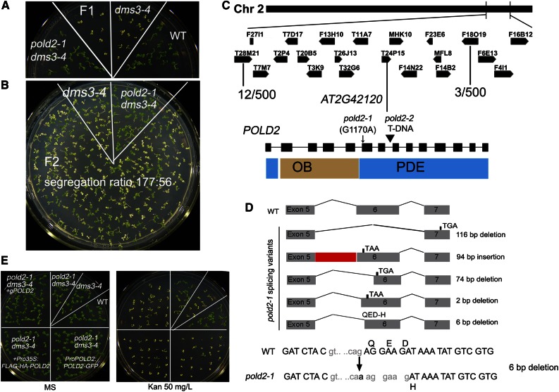
Figure 2.
Map-based cloning of POLD2. A, Phenotypic analysis of F1 seedlings of dms3-4 pold2-1 backcrossed with dms3-4 on kanamycin-containing medium. B, The segregation of F2 seedlings growing on kanamycin-containing medium. C, Map-based cloning of the pold2-1 mutation. The position of the POLD2 mutation was narrowed to the bottom of chromosome 2 between BAC T28M21 and F18O19. The mutation of pold2-1 occurred at splicing sites (G1170A). pold2-2 is a T-DNA insertion allele. The domain structure of the POLD2 protein is indicated. OB, oligonucleotide-/oligosaccharide-binding domain; PDE, phosphodiesterase-like domain. D, Spliced forms of cDNA caused by the pold2-1 mutation. Five kinds of transcripts were identified from 23 independent clones amplified from cDNAs. Among them, four transcripts would produce an earlier stop codon, and one would delete 6 bp and lead to the deletion of two amino acids (delete QE) and to the mutation of one amino acid (from original D to H). E, Complementary analyses of pold2-1 with genomic DNA or Pro35S:FLAG-HA-POLD2 construct, or native promoter driving cDNA (ProPOLD2:POLD2-GFP). Phenotypes of mutant and complementary lines growing on MS medium containing 0 mg/L kanamycin (left) or 50 mg/L kanamycin (right).

