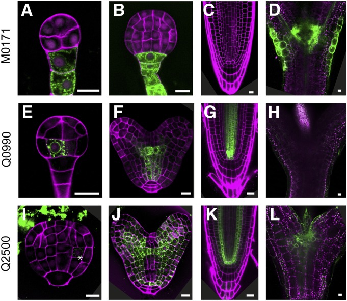
Figure 1.
GFP expression in early embryogenesis lines. GFP fluorescence in preglobular or globular stage embryos (A, E, and I), late globular or heart stage embryos (B, F, and J), root tips (C, G, and K), and shoot apex (D, H, and L) of M0171 (A–D), Q0990 (E–H), and Q2500 (I–L) lines. Magenta counterstaining in A, B, E, F, I, and J is Renaissance fluorescence, propidium iodide in C, G, and K, and chlorophyll autofluorescence in D, H, and L. Bars = 10 μm.

